TAVI/TAVR Procedure in Mumbai

The TAVI or TAVR procedure can be done through very small openings that leave all the chest bones in place. Transcatheter Aortic Valve Implantation (TAVI) also known as Transcatheter Aortic Valve Replacement (TAVR) is advised to the patient to take up this procedure when the adult is fragile to take the trauma of an open-heart surgery. A person’s experience with a TAVR procedure can be rated similar to a coronary angiogram in terms of recovery. Post the procedure it is likely spend less time in the hospital after TAVR as compared to a surgical valve replacement. Risk levels may be the same but the recovery is quicker because it is less invasive as compared to open heart surgery.
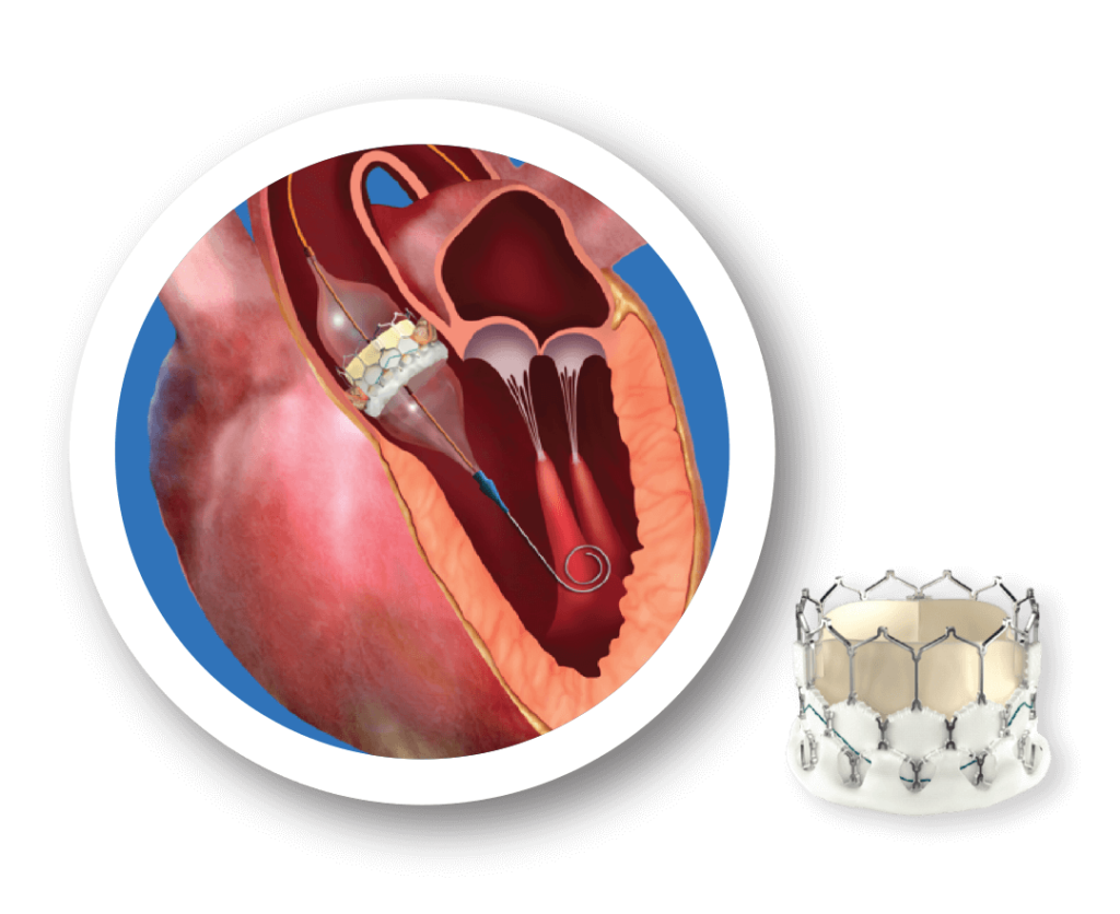
TAVI as the name suggests is a process wherein through the catheter an aortic valve is implanted or replaced. Doctors use a catheter (a long, flexible tube that doctors use to reach parts of the body that are usually hard to access, like blood vessels or the heart) to go through a blood vessel, often in the leg or groin, to reach the heart and insert a new valve.
- To explain the procedure simply, imagine your heart is like a high-traffic highway with an old, worn-out toll booth (the aortic valve) that cars (blood) have to pass through. Over time, the toll booth gets rusted and stuck & slow moving, making it harder for cars to get through, which causes a traffic jam (poor blood flow). Instead of closing down the whole highway to replace the toll booth (which would be like open-heart surgery), you use a clever method to add a brand new, fully functional toll booth right over the old one.
A team of workers (doctors) bring in a long, flexible arm (the catheter), which slides through the roadways (your blood vessels) to place the new booth in position. Once it’s set up, the new toll booth opens smoothly, letting the cars flow freely again, and the traffic jam clears up — all without any major construction or shutting down the highway.
- TAVI is minimally invasive procedure which means faster recovery, less complications compared to surgery
- The procedure is done by giving a general anaesthesia or conscious sedation
- The length of the procedure is usually 1-2 hours
The TAVR Procedure is Performed using Two Approaches
Transfemoral Approach: This is an approach of entering into the body without requiring a surgical incision in the chest. The invasion is done through the femoral artery which is a large artery in the groin.
Transapical Approach: With this approach, doctor enters into the body with a small incision in the chest through either a large artery in the chest or through the tip of the left ventricle which is also called as the apex.
Step By Step Procedure
1) The procedure begins with a small incision in the groin (in case of femoral approach).
2) After creating an opening in a blood vessel called the femoral artery doctor inserts a flexible tube, called an introducer sheath.
3) Doctor then inserts a flexible guidewire into the femoral artery (most preferred) through the sheath.
4) The guidewire is then passed through the femoral artery up to aorta and is pushed through the opening in the aortic valve into the left ventricle.
5) Doctor uses the guidewire to pass a catheter through the aortic valve. Then a balloon on the tip of the catheter is inflated to widen the opening in the valve. This is done to push the valve leaflets to the sides.
6) The replacement valve is compressed and placed on the tip of another catheter. This catheter is inserted through the aortic valve.
7) Once the catheter reaches the opening in aortic valve, a balloon is inflated underneath the replacement valve to expand it and the stent.
8) Post the placement of the new aortic valve, the doctor deflates the balloon.
9) In expectation that the stent will support and secure the replacement valve in place, the catheter and guidewire is removed.
10) At the end, the introducer sheath in the groin is removed and the skin incision is closed.
Note: There will be another incision on the other side of the groin through which instruments are inserted to monitor the heart during the procedure. This skin incision is also closed after the procedure. Insertion of the flexible tube catheter is well guided using advanced imaging technology like ultrasound or x-ray to carefully reach the aortic valve and deliver the new valve over the old.
Different types of valves used in TAVI:

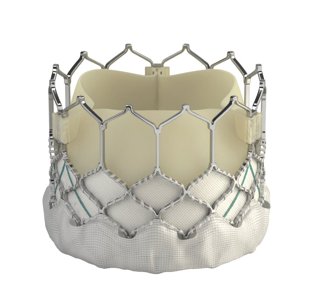
Balloon Expandable Valve

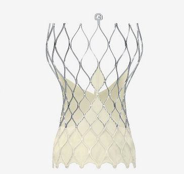
Self-Expanding Valve
Balloon-expandable valves: Balloon inflation is necessary to deploy and secure these valves, which are attached to a balloon catheter. The stent frame is expanded and the valve is released by clamping the valve onto a balloon, which is then inflated inside the native valve annulus.
Self-expanding valves: the stent frame of these valves is made of nitinol, a shape-memory alloy, which expands without the use of a balloon. The valve is put in a collapsed form into a catheter. When it reaches its destination, it is released, and the stent’s self-expanding characteristics enable it to open and secure itself inside the aortic annulus.
Key Differences Between Balloon-Expandable and Self-Expanding Valves
| Feature | Balloon-Expandable Valves | Self-Expanding Valves |
|---|---|---|
| Stent Frame Material | Stainless steel or cobalt-chromium | Nitinol (shape-memory alloy) |
| Deployment Method | Balloon inflation | Self-expands upon release |
| Precision of Placement | High (due to balloon control) | Adjustable/repositionable |
| Radial Force | Higher initial force | More gradual, gentle force |
| Anatomical Suitability | Best for calcified or small annuli | Better for larger or irregular annuli |
| Risk of Annular Rupture | Higher due to balloon force | Lower due to gradual expansion |
Who should opt for TAVI?
The ideal candidate for TAVI is someone whose body cannot withstand the trauma of open-heart surgery. In addition, a patient with severe aortic stenosis who is weak or has additional health conditions that make surgery more perilous, such as diabetes or hypertension. Because TAVI employs a replacement valve, candidates will not require anticoagulants for more than a few weeks following surgery. However, for the rest of one’s life, one may have to take blood thinners such as aspirin or clopidogrel.
Symptoms:
Aortic valve stenosis is hard to detect because the symptoms start to usually show when the condition gets severe with the heart valve getting narrower. It is important to consult a doctor when one notices the following frequently
- Shortness of breath
- Chest pain, discomfort or angina
- Dizziness or fainting
- Swelling in the legs and or abdomen
- Tiredness when you are increasing your activity level
- Heart palpitations (rapid, fluttering heartbeat)
- Congestive heart failure
It should be noted that the symptoms may be outcome of any other underlying condition hence the patient should consider complete evaluation from a cardiologist to ensure correct diagnosis.
Role of the heart team (cardiologist, cardiac surgeon, anaesthetist) becomes paramount in evaluating patients, diagnosing the actual condition and offer specialised treatment.
Preparing for TAVI
After going through series of tests and getting a confirmation with doctors about Aortic Stenosis and preparing for TAVI procedure one must be cautious of the following:
1) Look after oral (mouth) health:
- A dental check-up before having the TAVI procedure is important.
- Make an appointment with the dentist, if one hasn’t had one in the last 6 months
- If one needs teeth removed or treatment for gum disease, this must be done before the TAVI procedure
2) Learn more about the process
- Collate all the questions and email or call the office of the institution opted to review with one of the clinical coordinators
- Ensure that if you have an accompanying care giver, he/she is well acquainted with all the information and has the important numbers in case of running a query or reaching out
Pre-operative evaluations:
Apart from the other tests echocardiogram, CT scan, blood tests, and risk assessment are the most important evaluation tests before the procedure.
1) Echocardiography, commonly referred to as an echo, is a non-invasive imaging technique that uses ultrasound waves to create detailed images of the heart. It is one of the most widely used diagnostic tools in cardiology. Echocardiography is an essential tool for diagnosing, guiding, and monitoring cardiac conditions, particularly useful in interventions like TAVI due to its real-time imaging capabilities and detailed visualization of heart structures. There are two types of echo used in TAVI.
- Transthoracic Echocardiogram (TTE) Provides images of the heart chambers, valves, and major blood vessels.
- Transoesophageal Echocardiogram (TEE) Commonly used during surgical or catheter-based procedures (e.g., TAVI) for real-time monitoring. Echo is vital in all the three stages of the procedure, pre, during and post-surgery.
2) A CT scan, or Computed Tomography scan, is a medical imaging technique that uses X-ray beams and computer processing to create detailed cross-sectional images of the body. It provides highly accurate, three-dimensional images, making it a great tool for diagnosing and assessing a wide range of illnesses, especially in cardiology and vascular medicine.
These scans are important for assessing cardiovascular structures with high precision, making them an essential resource for organizing and enhancing results for procedures like TAVI. The scan helps in the pre-evaluation stage of the procedure because it ensures accurate measurement of the aortic valve and annulus for proper valve sizing. It assesses the peripheral vascular system to determine the safest access route and provides a comprehensive 3D view of the heart and its surrounding structures, reducing the risk of complications during the procedure.
Here is a comprehensive list of the tests and their purpose.
| Evaluation | Purpose |
|---|---|
| Clinical Assessment | Assess symptoms, comorbidities, and frailty |
| Echocardiography (TTE/TEE) | Evaluate aortic valve structure and function |
| CT Angiography | 3D assessment of aortic root, annulus, and access route |
| Electrocardiogram (ECG) | Identify arrhythmias and conduction issues |
| Pulmonary Function Tests | Assess lung function and respiratory risk |
| Blood Tests | Check renal function, coagulation, and overall health |
| Geriatric and Frailty Assessment | Evaluate functional status in elderly patients |
| Heart Team Consultation | Multidisciplinary decision-making |
| Anaesthetic Evaluation | Plan anaesthesia approach and manage risks |
Additionally, as a candidate for the surgery one must consult with regards to medications that need to be adjusted or stopped before the procedure. And the date of hospitalisation and the admission process and what to expect on the day of the procedure.
Recovering in hospital after your TAVI
Recovery in the hospital is taken care by the health providers. The nurse checks the patient regularly to make sure he/she is comfortable. The nurse regularly checks incision areas, guides on what to expect as one heals and how to care for these sites. The nurse reminds the patient to keep their legs straight the first 4 hours after the procedure. This is very important to prevent bleeding from the insertion sites in the groin. Also, there may be some intravenous tubes or temporary wires attached. After the first 4 hours, the nurse helps the patient to gradually increase the activity, from sitting up in bed to sitting in a chair to walking short distances. When one is fully awake, the institute provides the patient clear fluids to drink. One can start eating food once one starts drinking well.
| What is normal? | What are warning signs? |
|---|---|
| Slightly red, swollen and tender | Any area of redness or swelling that does not get better |
| A bit lump or bumpy | A lump that is getting bigger |
| Occasional clear drainage | Yellow or pink drainage |
| Pain gets better gradually | Pain does not get better or is severe |
| Bruising | Fever – a temperature of 38.5 °C(101 °F) or higher |
| Bleeding from the puncture site that does not stop |
Benefits of TAVI
- Minimally invasive, lower risk compared to open-heart surgery
- Shorter recovery time and reduced hospital stay
- Suitable for high-risk patients
- Improvement in symptoms like breathlessness, fatigue, and chest pain
Risks and Potential Complications
Valve surgery is generally very successful, and it can have many benefits. It can help to relieve symptoms, or get rid of them completely. It could help you live longer and improve your quality of life. But, like any other medical procedure, there are risks:
Patients undergoing TAVI have risk
- Develop arrhythmias and need for a pacemaker
- Get bleeding
- Get a valve leakage (paravalvular leak)
- Other vascular complications
- Age
- Current state of health
- If one is getting a valve replaced or repaired
- The level of patients’ condition
- Whether you’re having any other procedure simultaneously
- Which valve is affected
While the risk for above adverse events is minimal, a patient should always remember that with development in medical technologies, the benefits of TAVI outweigh the anticipated risks. It is also to be noted that with developing aortic stenosis condition, patient is susceptible to various issues and hence should consult experienced cardiologist team.
Comparison Between TAVI and Surgical Aortic Valve Replacement (SAVR)
Comparison of TAVI vs. traditional open-heart aortic valve replacement can be seen through the following parameters:
- Invasiveness
- Recovery time
- Risks and benefits for different patient groups
Long-term Outcomes of TAVI
- Improvement in quality of life and symptom relief
- Longevity of the replacement valve (how long the valve typically lasts)
- Importance of follow-up visits to monitor valve function and overall heart health
What do I need to do after I am discharged from hospital?
Once discharged from the hospital one must be mindful of the things one can and cannot do.
List of Don’ts:
- Think clearly. Surgery and pain-management medication can often hamper your thinking skills, so do not make important decisions during this time
- Easily sit up from a lying position
- Raise your arms overhead for dressing
- Prepare a meal
- Get your incision wet
- Lift a jug of milk
- Tie your shoes
- Climb the staircase
- Run the vacuum
- Carry a bag
List of Do’s:
- Follow your doctor’s directions
- Plan your nutrition
- Get rest and accept help
- Plan your environment and reread your discharge instructions
- Verify all medications. List and track your doses too
- Ensure that one has all the important numbers handy
Get in Touch!
Address: A12 Silver Apts, Shankar Ghanekar Rd, Behind Siddhivinayak Mandir, Prabhadevi, Mumbai, Maharashtra 400025
Phone: +91-9004506263
