What is Ventricular Septal Defect? Types, and Treatment Options

When a parent learns their newborn has a heart defect, the news can be terrifying. It can be just as overwhelming for an adult to receive the same diagnosis after years of inexplicable symptoms. The condition behind both situations is often the same: a ventricular septal defect, one of the most common structural heart conditions diagnosed today.
The reassuring truth is that not every ventricular septal defect requires immediate treatment, and when it does, the options available today are safer, more precise, and far less invasive than most people expect.
This blog walks you through everything you need to know in plain language, without the medical jargon.
What Is a Ventricular Septal Defect?
To understand a ventricular septal defect, it helps to picture how the heart is built. Your heart has four chambers: two on the top (the atria) and two on the bottom (the ventricles). The left ventricle pumps oxygen-rich blood out to the body, while the right ventricle pumps oxygen-poor blood to the lungs to be refreshed. A thick wall called the septum separates the two ventricles, keeping these two flows of blood completely apart.
A ventricular septal defect or VSD, is simply an opening or hole in this dividing wall. When this hole is present, oxygen-rich blood from the left side leaks across to the right side, mixing with oxygen-poor blood. This forces the heart and lungs to work harder than they should to maintain normal circulation. Over time, if left unaddressed, this extra workload can strain the heart muscle and affect the lungs.
VSD is one of the most common congenital heart diseases, with a prevalence of approximately 4 to 6 per 1,000 live births. However, it is not exclusively a condition of childhood; VSDs can also develop in adults, sometimes as a complication of a heart attack or heart surgery.
Types of Ventricular Septal Defect
Not all VSDs are the same. They vary in size, location, and clinical significance. Understanding the type helps determine the most appropriate treatment approach.
- Perimembranous VSD – This is the most common type, accounting for the majority of clinically significant cases. It occurs in the upper part of the ventricular septum, near the heart valves.
- Muscular VSD – Located in the lower, muscular portion of the septum, these defects are surrounded entirely by muscle tissue.
- Inlet VSD – Found near the inlet valves of the ventricles, this type is less common and is sometimes associated with other congenital heart conditions.
- Outlet VSD – Also called supracristal or conal VSDs, these are located near the outflow tracts of the ventricles where blood exits the heart.
The size of the defect is as important as its location. Small defects may cause no symptoms and close without any treatment. Large defects, on the other hand, can significantly affect heart and lung function and require timely management.
How Is a Ventricular Septal Defect Diagnosed?
Diagnosis typically begins with a clinical examination where a doctor detects a characteristic heart murmur. From there, the key investigations include an echocardiogram, an ultrasound of the heart that shows the location, size, and direction of blood flow through the defect. This is the most important diagnostic tool and provides the information needed to plan treatment.
An ECG and chest X-ray may also be performed to assess any impact on the heart’s size and electrical function. In more complicated cases, an MRI or cardiac catheterisation may be recommended for a more detailed assessment.
At Heart Valve Experts, we use advanced 3D echocardiography for precise diagnosis and treatment planning, ensuring every decision is tailored to the individual patient’s anatomy and condition. You can read more about how structural heart conditions are diagnosed and treated on our website.
Ventricular Septal Defect Treatment Options
Treatment for a VSD depends on its size, type, location, and the symptoms it is causing. Not every VSD requires intervention. Here is an overview of the options available:
Watchful Waiting
For small defects, particularly muscular VSDs in infants, the recommended approach is careful monitoring with regular echocardiograms. Many small VSDs close naturally as the child grows. The child’s development, growth, and feeding are all closely monitored during this period.
Medication
Medications do not repair the defect itself but can help manage symptoms such as fluid retention, reduce the workload on the heart, and support a child’s growth while monitoring whether the defect closes on its own. The treatment is typically a bridge measure rather than a long-term solution.
Device Closure: Minimally Invasive
For eligible patients, VSD closure is a minimally invasive procedure where a small incision is made in the femoral vein or artery in the groin. A catheter is then guided to the heart using fluoroscopy and echocardiography, and a specialised VSD closure device is placed across the defect using a controlled release technique.
At Heart Valve Experts, our team routinely performs device-based VSD closure procedures using the latest equipment for accurate diagnosis and treatment. Learn more about VSD closure in Mumbai on our dedicated procedure page.
Surgical Repair
When the defect is large, located in a position that is not accessible by catheter, or associated with other cardiac abnormalities, open-heart surgical repair may be recommended. A patch is used to close the hole indefinitely.
What Happens After Treatment?
Recovery after device closure is generally swift. Recovery after ventricular septal defect surgery typically takes 4 to 6 weeks, with shorter recovery times for catheter-based closures. Patients are given medications for a while after the procedure to help them heal and avoid problems. Regular follow-up appointments and echocardiograms are scheduled to confirm the device is in position and the heart is functioning well.
For most patients, a successfully treated VSD allows them to return to a full, active, and unrestricted life. Many children who undergo treatment go on to lead completely normal lives with no long-term limitations.
Conclusion
A ventricular septal defect can feel like a frightening diagnosis, but it’s also one of the most treatable structural heart conditions available in modern cardiac care. Whether the right approach is monitoring, device closure, or surgery, outcomes today are better than ever. The most important step is getting the right evaluation from a team that genuinely understands structural heart conditions.
If your child has been diagnosed with a ventricular septal defect (VSD), or if you are an adult living with a previously identified but unresolved VSD, it is important not to leave this condition without proper evaluation and clarity. Book a consultation with Heart Valve Experts, Mumbai, and get a clear path forward.
FAQs
In some cases, yes. Small muscular VSDs in infants and young children may close naturally as the heart grows. Your cardiologist will monitor these events closely with regular echocardiograms.
Device closure is minimally invasive and performed under sedation, so patients experience minimal discomfort.
Yes. Some small VSDs go undetected through childhood and are picked up incidentally in adulthood. VSDs can also develop in adults as a rare complication of a heart attack or cardiac surgery.
A small, asymptomatic VSD may require monitoring but not treatment. A large, untreated VSD can over time lead to complications including pulmonary hypertension and heart failure. Your cardiologist will advise based on your specific situation.
Heart Valve Experts in Mumbai specialise in minimally invasive device closures for ventricular septal defects. Our team offers a thorough evaluation, advanced imaging, and personalised treatment plans.

About The Author
Dr. Ankur U. Phatarpekar M.D., D.M., FSCAI
With over 15 years of expertise, Dr. Phatarpekar is recognised as a renowned interventional cardiologist in Mumbai, specialising in complex coronary interventions, structural heart procedures, and pioneering work in Transcatheter Aortic Valve Implantation (TAVI).
● Blog
Related Heart Valve Experts (HVE) Blog
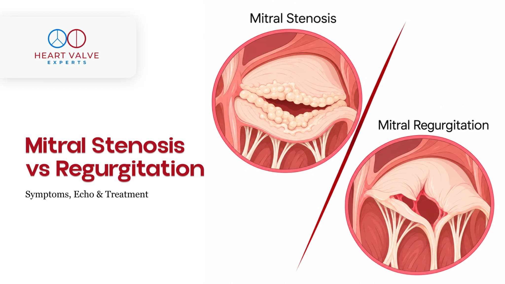
Mitral Stenosis vs Mitral Regurgitation: Differences in Symptoms, Echo Findings and Treatment
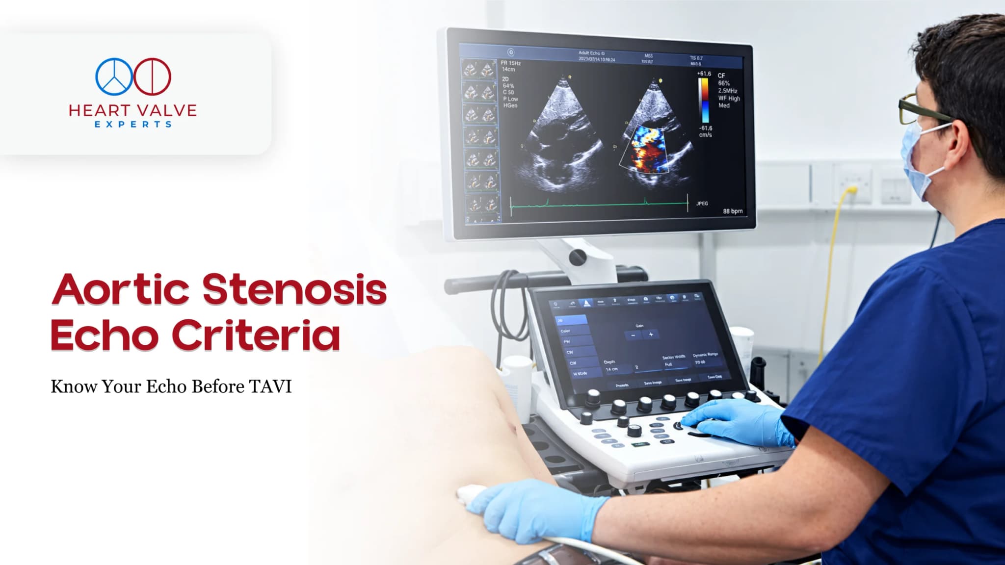
Aortic Stenosis Echo Criteria Explained: Understanding Your Echo Before a TAVI Assessment
How to Get a Medical Second Opinion for Heart Valve Treatment Before Travelling to India
Medical Visa for India: Requirements and Application Guide for Heart Patients
Heart Valve Surgery in Mumbai: How to Choose a Hospital and When to Seek a Second Opinion