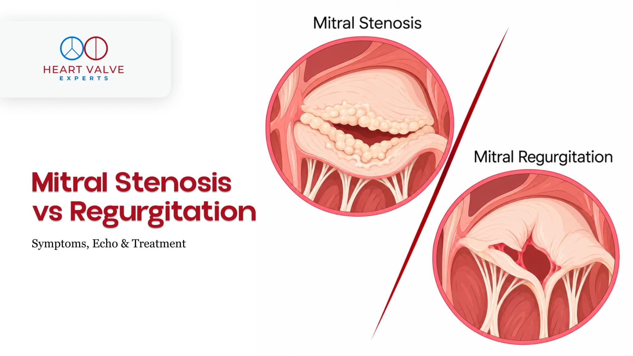
If your echocardiogram mentions a mitral valve problem, you may see the terms mitral stenosis or mitral regurgitation.
They affect the same valve in different ways. Mitral stenosis means the valve does not open fully, making it harder for blood to move forward. Mitral regurgitation means the valve does not close properly, allowing some blood to leak backward. Some people can have both problems.
Symptoms such as breathlessness or tiredness can occur with either condition, so symptoms alone cannot tell you which valve problem you have. Your cardiologist will consider your echo findings, symptoms, heart rhythm and how the condition is affecting your heart.
This guide explains the main differences and what the terms on your report may mean. It does not replace interpretation by your cardiologist.
The mitral valve sits between the left atrium, the heart’s upper-left chamber, and the left ventricle, its main pumping chamber.
In mitral stenosis, the valve opening becomes narrowed, making it harder for blood to move into the left ventricle. This can increase pressure behind the valve and affect the lungs.
Worldwide, rheumatic heart disease remains an important cause of mitral stenosis. In older adults, calcium can also build up around the mitral valve and make it harder for the valve to open.
In mitral regurgitation, the mitral valve does not close tightly. When the left ventricle contracts, some blood moves forward into the aorta, while some leaks backward into the left atrium.
Over time, significant leakage can enlarge parts of the heart and increase pressure in the lungs.
Mitral regurgitation is often described as primary or secondary.
Primary mitral regurgitation is caused by a problem with the valve or its supporting structures, such as mitral valve prolapse or degenerative valve disease.
Secondary mitral regurgitation occurs when changes in the left ventricle, left atrium or surrounding valve ring prevent the valve from closing properly.
The distinction is important because the cause of the leakage can affect which treatment options are considered.
A simple way to remember the difference is:
| Feature | Mitral stenosis | Mitral regurgitation |
| Main valve problem | Valve opening is too narrow | Valve does not close properly |
| What happens to blood flow? | Blood has difficulty moving from the left atrium to the left ventricle | Some blood leaks backward into the left atrium |
| Common causes | Often rheumatic valve disease worldwide; calcium build-up can also cause narrowing | May result from disease of the valve itself or changes in the heart that affect valve closure |
| What does the echo assess? | Valve area, pressure gradient, valve anatomy and pressure in the lungs | Severity and cause of the leak, heart chamber size and heart function |
| Possible procedures | Balloon commissurotomy in suitable rheumatic cases or surgery when needed | Valve repair or replacement, with TEER or other transcatheter treatment in selected patients |
| Can both occur together? | Yes | Yes |
This table is a simple comparison, not a diagnostic tool. Your cardiologist will interpret your echo findings together with your symptoms and other clinical information.
There is a lot of overlap.
As mitral stenosis becomes more significant, pressure can build up in the left atrium and lungs. Symptoms may include shortness of breath, tiredness and reduced ability to exercise.
Some people may also develop an irregular heart rhythm called atrial fibrillation or increased pressure in the blood vessels of the lungs.
Mild mitral regurgitation may cause no symptoms.
When the leakage becomes more significant, symptoms can include shortness of breath, tiredness or weakness, light-headedness and palpitations. Some people may feel more breathless when lying flat or wake during the night feeling short of breath.
Because both conditions can cause breathlessness, fatigue and palpitations, symptoms alone cannot reliably tell whether you have mitral stenosis or mitral regurgitation.
The causes are different, although some conditions can affect the mitral valve in more than one way.
For mitral stenosis, rheumatic heart disease remains an important cause worldwide. Calcium build-up around the valve can also cause narrowing, particularly in older adults.
The causes of mitral regurgitation are more varied. The problem may come from the valve itself, or from changes in the heart that prevent the valve from closing properly.
This distinction matters because treatment for secondary mitral regurgitation often includes treating the underlying heart condition before deciding whether a valve procedure is needed.
They often are, but a murmur alone cannot tell how severe the valve problem is.
Mitral stenosis typically causes a low-pitched murmur during the part of the heartbeat when the heart is relaxing and filling. An additional sound called an opening snap may also be heard.
Mitral regurgitation usually causes a murmur while the left ventricle is contracting, often heard most clearly near the apex of the heart.
Heart sounds can vary depending on the valve problem, heart rhythm and other clinical factors. Your doctor will therefore use the examination together with an echocardiogram and other findings rather than relying on the murmur alone.
An echocardiogram, or echo, uses ultrasound to look at the mitral valve and heart chambers.
In mitral stenosis, the echo assesses how well the valve opens, the size of the opening, the pressure gradient across the valve and how the narrowing is affecting the heart and lung circulation. In rheumatic disease, the valve leaflets may appear thickened or have restricted movement.
In mitral regurgitation, the echo assesses how much blood is leaking backward, why the leak is occurring and how it is affecting the left atrium and left ventricle. It can also help identify problems such as mitral valve prolapse or a flail leaflet.
A standard transthoracic echo is usually the first test. Sometimes a more detailed transoesophageal echocardiogram (TOE/TEE) or other imaging is needed if the first study does not provide enough information or if a valve procedure is being planned.
Neither mitral stenosis nor mitral regurgitation is graded from one number alone.
For rheumatic mitral stenosis, a mitral valve area of 1.5 cm² or less is commonly considered within the severe range. Doctors also consider your symptoms, pressure across the valve, heart rhythm and how the narrowing is affecting the heart and lungs.
For mitral regurgitation, severity is assessed using several echo findings. These include how much blood is leaking backward, the cause of the leak, heart chamber size and heart function.
Your cardiologist will also consider whether:
These findings are considered together when deciding whether treatment is needed and which options may be appropriate.
Medicines can help manage symptoms and related heart problems, but they do not physically correct a narrowed or leaking valve.
For mitral stenosis, medicines may help reduce fluid build-up, control the heart rate or rhythm, and lower the risk of blood clots in selected patients.
People with rheumatic mitral stenosis and atrial fibrillation may need anticoagulation to reduce the risk of stroke or other blood clots. The type of anticoagulant is important in this setting and needs individual medical guidance. Do not start, stop or change an anticoagulant without advice from your treating clinician.
For mitral regurgitation, medicines may help manage heart failure, high blood pressure, fluid retention or rhythm problems, depending on the cause.
In secondary mitral regurgitation, treating the underlying heart condition is often an important part of management before a valve procedure is considered.
For some people with rheumatic mitral stenosis, a catheter procedure can open the narrowed valve without replacing it surgically.
This treatment may be called balloon mitral valvotomy, percutaneous mitral balloon commissurotomy (PMBC) or percutaneous mitral commissurotomy (PMC). A balloon is passed through a catheter and inflated across the valve to separate fused leaflet edges.
It is not suitable for everyone. Specialists consider the valve anatomy, the amount of mitral regurgitation and whether a blood clot is present in the left atrium.
For selected people with symptomatic severe rheumatic mitral stenosis, suitable valve anatomy, no left-atrial thrombus and less than moderate mitral regurgitation, balloon commissurotomy can be an important treatment option. Surgery may be considered when the anatomy or other clinical factors make the catheter procedure unsuitable.
Treatment depends on whether mitral regurgitation is primary or secondary, how severe it is, your symptoms, heart function, valve anatomy and overall health. Some people need regular monitoring, while others may need medicines or a valve procedure.
For severe primary degenerative mitral regurgitation, surgical repair is often preferred when a lasting repair is likely. If a procedure is being considered, understanding the difference between mitral valve repair and replacement can help you discuss the options with your specialist.
Transcatheter edge-to-edge repair (TEER) is a catheter-based procedure that brings parts of the mitral valve leaflets together to reduce mitral regurgitation. MitraClip is one of the devices used for this type of treatment.
TEER and MitraClip can be considered for selected people with severe mitral regurgitation. This includes some patients with symptomatic primary MR who have a high surgical risk and suitable valve anatomy.
TEER may also be an option for selected people with severe secondary MR who continue to have symptoms despite treatment for the underlying heart condition.
Transcatheter mitral valve replacement (TMVR) aims to replace the mitral valve using a catheter-based approach rather than conventional open surgery.
TMVR is a highly specialised treatment and is different from TEER. Whether it may be suitable depends on the type of mitral valve disease, valve anatomy, previous procedures, the amount and location of calcium, and other clinical factors.
For selected patients with severe mitral valve disease and extensive mitral annular calcification, a transcatheter approach may be considered at an experienced Heart Valve Centre.
An echo is an important part of the assessment, but it cannot by itself determine whether someone is suitable for TMVR, TEER, balloon valvotomy or surgery.
Yes. A mitral valve can sometimes be both narrowed and leaky. This is called mixed mitral valve disease.
When both problems are present, your cardiologist looks at which one is having the greater effect and how the combination is affecting blood flow, the heart and the lungs.
If your echo report mentions both mitral stenosis (MS) and mitral regurgitation (MR), the findings need to be interpreted together rather than relying on a single severity label.
If you are in Bangladesh and considering a specialist review in India, send the records you already have before making travel plans. Your latest echocardiogram report and a brief description of your symptoms are a good starting point. You can also include previous echo reports, recent cardiology notes, an ECG, your current medicines and details of any earlier heart procedures.
If you have already had a transoesophageal echo, CT scan or coronary angiogram, share those reports if requested. You do not need to arrange new tests just to make an enquiry. After reviewing your records, the specialist can advise whether anything else is needed.
If travelling to Mumbai becomes an option, you can read about planning your visit from Bangladesh. A review of your records can help identify possible next steps, but a final treatment decision may require an examination and further tests.
Button: Request an Expert Opinion
A remote review can help clarify your existing findings and possible next steps. It does not by itself confirm a final diagnosis, treatment choice or eligibility for a particular procedure.
Do not wait for an international consultation if you suddenly become unwell.
Seek urgent local medical care for severe or rapidly worsening shortness of breath, new or severe chest pain, fainting, coughing up a significant amount of blood, signs of a stroke, or any sudden major deterioration.
If you develop worsening breathlessness, swelling, palpitations, reduced ability to exercise or new dizziness, arrange prompt assessment with your local treating clinician.
Neither is automatically worse. Both conditions can range from mild to severe. What matters most is how severe the valve problem is, whether you have symptoms, and how it is affecting your heart and lungs.
Usually, yes. An echocardiogram can show whether the mitral valve is narrowed, leaking or both. It also provides measurements that help assess severity and how the valve problem is affecting the heart.
No. Rheumatic heart disease is a common cause worldwide, but mitral stenosis can also result from calcium build-up around the valve, particularly in older adults, and less commonly from congenital or other conditions.
Sometimes. Mild mitral regurgitation may only need monitoring. In secondary mitral regurgitation, treating the underlying heart condition can help reduce symptoms and may improve the leakage. Selected patients with severe MR may also be considered for catheter-based treatments such as TEER.
No. Balloon mitral valvotomy is used for selected patients with rheumatic mitral stenosis, where the valve opening is narrowed. It does not treat isolated mitral regurgitation. Significant MR can also make balloon treatment unsuitable in someone who has mitral stenosis.
TEER reduces mitral regurgitation by bringing parts of the existing valve leaflets closer together. TMVR replaces the mitral valve using a catheter-based procedure. They are different treatments used in different situations, and suitability depends on the type of valve disease, anatomy, heart function and other clinical factors.

About The Author
With over 15 years of expertise, Dr. Phatarpekar is recognised as a renowned interventional cardiologist in Mumbai, specialising in complex coronary interventions, structural heart procedures, and pioneering work in Transcatheter Aortic Valve Implantation (TAVI).
● Blogs