If your echocardiogram shows aortic stenosis, you may see terms such as peak velocity, mean pressure gradient, aortic valve area and ejection fraction. These measurements help assess how narrow the valve is and how your heart is functioning.
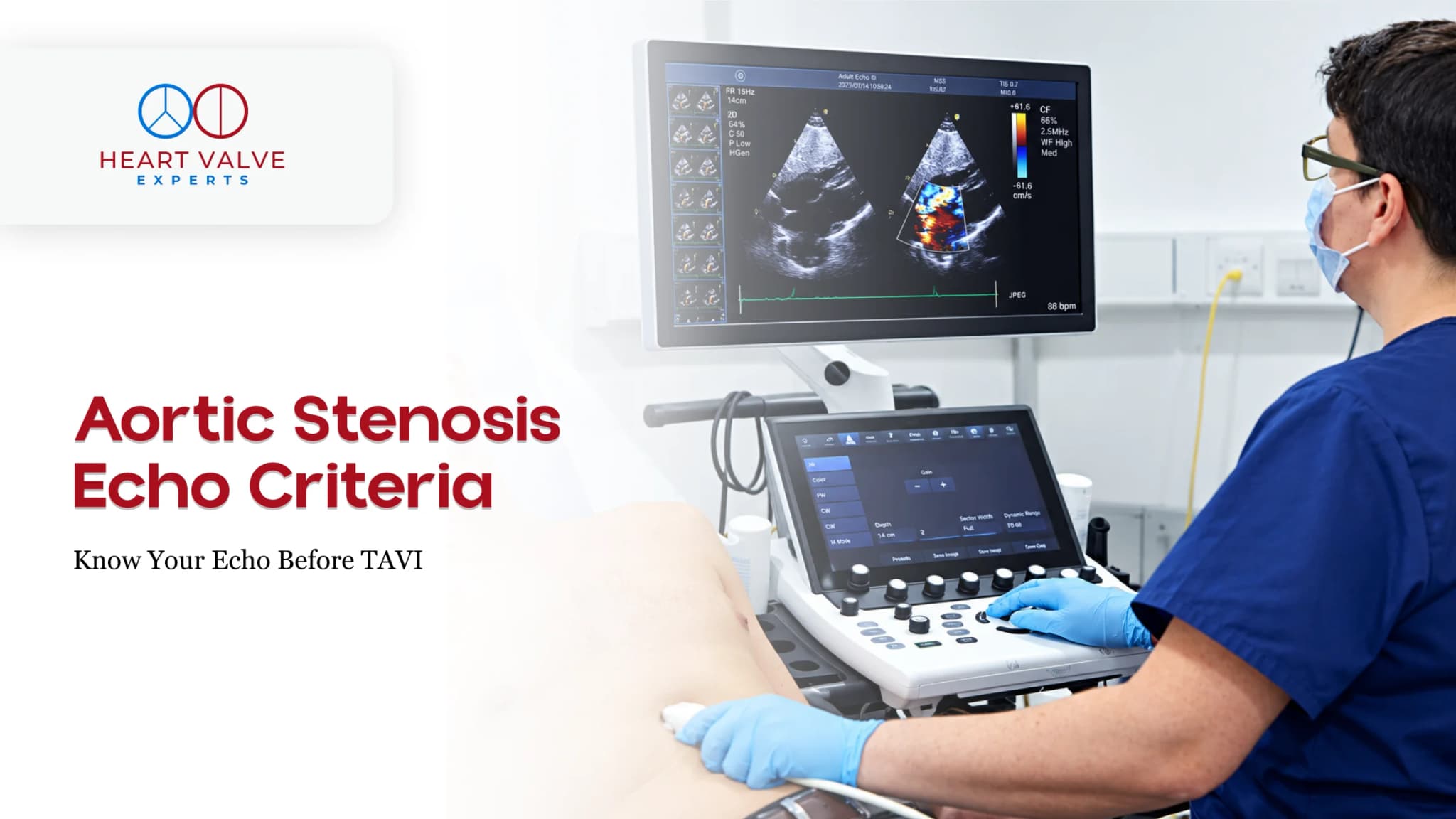
An echo is the main non-invasive test used to assess aortic stenosis. Cardiologists look at several measurements together because one number alone may not reflect the full picture.
This article explains the main echo terms and what they may mean before a specialist or TAVI assessment. It cannot confirm how severe your condition is or whether TAVI is suitable for you.
The aortic valve sits between the heart’s main pumping chamber, the left ventricle, and the aorta, the large artery that carries blood to the body.
In aortic stenosis, the valve becomes narrowed, so the heart has to pump blood through a smaller opening.
An echo can assess both the valve and how the heart is responding. Important findings may include:
Peak velocity, mean gradient and valve area are assessed together, rather than relying on a single measurement.
Your report may use the terms Vmax, peak aortic jet velocity or maximum velocity. This is measured in metres per second (m/s).
It shows how fast blood is moving through the aortic valve. When the valve becomes narrower, blood often has to move faster through the smaller opening.
Commonly used echo ranges are:
However, velocity is only one part of the assessment. If blood flow through the heart is reduced, significant aortic stenosis may be present even when the velocity is not very high. Your cardiologist will therefore interpret this measurement together with the rest of your echo and your clinical condition.
Another important measurement on your echo is the mean gradient, reported in millimetres of mercury (mmHg).
It reflects the pressure difference as blood passes through the narrowed aortic valve. In general, a narrower valve creates a higher gradient, although the amount of blood flowing through the valve also affects the result.
Commonly used ranges are:
Your report may also mention a peak gradient. This is different from the mean gradient. Cardiologists usually interpret the mean gradient together with peak velocity, valve area and the rest of the echo findings.
Aortic valve area, or AVA, estimates the size of the opening in the aortic valve. It is usually measured in square centimetres (cm²).
Unlike velocity and pressure gradient, a smaller valve area generally means a narrower valve.
Commonly used ranges are:
You may also see an indexed AVA, which adjusts the measurement for body size. An indexed AVA below about 0.6 cm²/m² can be associated with severe aortic stenosis.
AVA is only one part of the assessment. Your cardiologist will interpret it together with peak velocity, mean gradient, blood flow and the rest of your echo findings.
The table below explains commonly used echocardiographic ranges for aortic stenosis. It is not a self-diagnosis table. Cardiologists interpret these measurements together with your symptoms, blood flow, heart function and other findings.
| Echo measurement | Mild | Moderate | Severe range |
| Peak aortic velocity | 2.6–2.9 m/s | 3.0 to <4.0 m/s | ≥4.0 m/s |
| Mean gradient | <20 mmHg | 20 to <40 mmHg | ≥40 mmHg |
| Aortic valve area (AVA) | >1.5 cm² | 1.0–1.5 cm² | About 1.0 cm² or less |
| Indexed AVA | >0.85 cm²/m² | 0.60–0.85 cm²/m² | <0.60 cm²/m² |
| Velocity ratio | >0.50 | 0.25–0.50 | <0.25 |
These are commonly used echocardiography ranges. Measurements near the boundaries need clinical interpretation. If the values do not agree, your cardiologist may review the images, blood flow and other clinical information before deciding how severe the stenosis is.
Sometimes the measurements on an echo do not match. For example, the valve area may be in the severe range while the peak velocity or mean gradient is lower.
This does not always mean the test is wrong. Differences can occur because of blood flow, blood pressure or how the measurements were taken.
This is why aortic stenosis severity is assessed using several findings together rather than one number alone.
Your cardiologist may review the original echo images, heart function and blood flow, and decide whether further testing is needed.
Your echo may also report left ventricular ejection fraction (LVEF). This shows how well the heart’s main pumping chamber is pumping blood.
LVEF does not measure how narrow the aortic valve is, but it helps doctors understand how aortic stenosis may be affecting your heart.
Your cardiologist may also look at heart muscle thickness, chamber size, blood flow and other valve problems when assessing your condition.
Your echo results are only part of the assessment. Doctors also consider your symptoms and whether another condition could be causing them.
Symptoms of significant aortic stenosis can include:
Treatment decisions are not based on one echo measurement alone. Your specialist will consider your symptoms, valve measurements, heart function and overall health when discussing the next steps.
An echo is the main test used to assess aortic stenosis. If the results do not clearly show how severe the narrowing is, your specialist may recommend further testing, such as a cardiac CT calcium score or, in selected cases, a stress echo.
If TAVI is being considered, CT may also help your team assess the valve, nearby structures and blood vessels as part of planning for the procedure. Severe aortic stenosis on an echo does not automatically mean TAVI is suitable for you. Your specialist will advise which tests you need after reviewing your results and overall health.
If you are considering travelling to India for a TAVI assessment, sharing your existing records first can help the specialist understand your condition and identify what else may be needed.
You may be asked to provide:
Send the records you already have. Do not arrange CT, angiography or other tests just to complete this list. The reviewing specialist can advise whether further tests are needed.
If you are travelling from Nigeria, you can find more information about planning your visit. Patients travelling from elsewhere can learn more about visiting HVE from abroad.
CTA: Share your latest echocardiogram and symptom history for specialist review.
Button: Request an Expert Opinion
An echo showing severe aortic stenosis is an important finding, but it does not by itself determine whether TAVI is the right treatment.
TAVI (transcatheter aortic valve implantation) replaces the aortic valve using a catheter rather than open-heart surgery. Specialists also consider your symptoms, overall health, heart and valve anatomy, blood-vessel access and other clinical factors when assessing whether it is suitable.
This is why people with similar echo results may have different treatment options.
A remote specialist review can help clarify whether further TAVI assessment may be appropriate, but it cannot confirm TAVI suitability or predict the outcome of treatment.
Do not wait for an international appointment or remote TAVI review if you become suddenly unwell.
Seek urgent local medical care for severe or rapidly worsening breathlessness, new or severe chest pain, fainting, or a sudden major deterioration in your condition.
If your symptoms are not an emergency but are new or getting worse, such as increasing breathlessness, chest discomfort, dizziness, fainting episodes or reduced ability to carry out normal activities, arrange prompt review with your local healthcare team.
International travel planning should never delay urgent or necessary medical care.
The main measurements are peak aortic jet velocity, mean pressure gradient and aortic valve area (AVA). Severe-range findings commonly include a peak velocity of 4.0 m/s or higher, a mean gradient of 40 mmHg or higher, and an AVA of about 1.0 cm² or less. These measurements are interpreted together, not in isolation.
Not always. An AVA below 1.0 cm² is in the severe range, but doctors also consider velocity, gradient, blood flow and other findings. If the measurements do not agree, further assessment may be needed.
A mean gradient of 40 mmHg or higher is in the severe range for aortic stenosis. However, it needs to be interpreted with the other echo measurements and your clinical condition.
No. An echo provides important information about your valve and heart, but TAVI suitability requires a broader assessment. Specialists also consider your symptoms, overall health, heart and valve anatomy, blood-vessel access and other clinical factors.
A CT scan may help clarify aortic stenosis when the severity remains uncertain. If TAVI is being considered, CT can also provide important information about the valve, surrounding anatomy and blood vessels used to reach the heart.
Not automatically. Send your most recent echo first and ask the reviewing specialist whether another study is needed. This can depend on how recent your echo is, whether your symptoms have changed and your current clinical condition.

About The Author
With over 15 years of expertise, Dr. Phatarpekar is recognised as a renowned interventional cardiologist in Mumbai, specialising in complex coronary interventions, structural heart procedures, and pioneering work in Transcatheter Aortic Valve Implantation (TAVI).
● Blogs