
Medically Reviewed By
People often use the phrase “heart blockage” to mean different things, and that confusion can make an already worrying situation feel even harder to navigate. If your doctor has mentioned a heart valve problem, it helps to understand exactly what kind of blockage they are referring to, how it gets confirmed, and what the findings mean for your next steps.
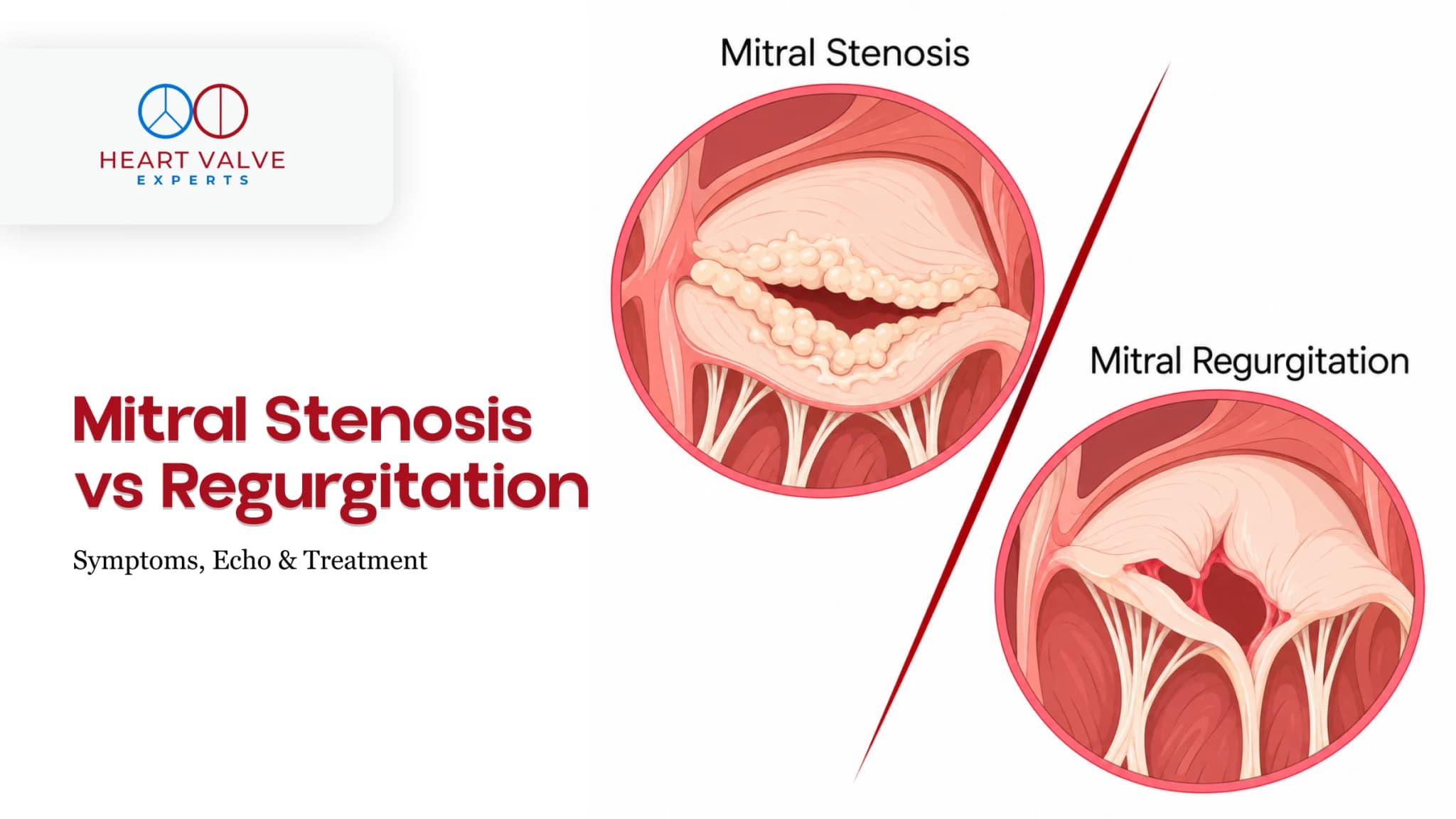
When people say heart valve blockage, they are usually describing valve stenosis, a condition where one of the heart’s valves has become narrowed, restricting blood flow. This is different from a coronary blockage, which refers to a buildup of plaque inside the arteries that supply blood to the heart muscle itself.
The two conditions can cause overlapping symptoms, which is why they are sometimes confused, but they are distinct problems with different diagnostic pathways and treatment approaches. Valve stenosis is a structural issue with the valve itself, most commonly affecting the aortic valve (between the left ventricle and the main artery) or the mitral valve (between the left atrium and left ventricle).
Knowing which type of blockage you are dealing with matters, because the tests and the treatment are not the same.
Heart valve blockage symptoms often build slowly, which means many people adjust to them without realising something is wrong. Common signs of heart valve blockage include:
In aortic stenosis specifically, the classic triad of symptoms is chest pain, breathlessness, and fainting. The appearance of any of these symptoms in someone with known valve disease is taken seriously, as it often signals that the condition has progressed to a point where intervention needs to be considered.
Some people have no symptoms at all and only discover a valve problem when their doctor listens to their heart and picks up a murmur.
A stethoscope is still one of the most valuable tools in detecting valve stenosis. A narrowed valve creates turbulent blood flow, which produces a characteristic sound called a heart murmur. Not all murmurs indicate disease, but certain murmur patterns are strongly associated with specific valve conditions and prompt further investigation.
If a murmur is detected, the next step is almost always imaging to confirm the diagnosis and assess severity.
Echocardiogram (Echo)
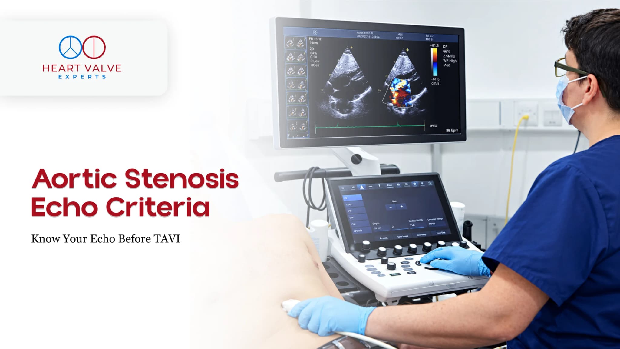
This is the primary test for diagnosing and grading heart valve stenosis. Using ultrasound waves, it produces real-time images of the heart’s structure and shows how blood moves through each valve. It measures the pressure gradient across the valve and the valve’s opening area, both of which determine how severe the narrowing is. A transthoracic echocardiogram (TTE) is the standard first-line test. A transoesophageal echocardiogram (TOE), where a probe is passed into the oesophagus, gives a more detailed view when the standard echo is not conclusive or when planning a procedure.
ECG (Electrocardiogram)
An ECG records the electrical activity of the heart and can identify changes in heart rhythm or thickening of the heart muscle, both of which can develop as the heart works harder against a narrowed valve. It does not diagnose stenosis directly, but it adds important information to the overall picture.
Chest X-ray
A chest X-ray can show an enlarged heart, calcium deposits on the valve, or fluid in the lungs, all of which may point to long-standing valve disease affecting heart function.
CT Scan
A cardiac CT provides highly detailed images of the valve’s anatomy and the surrounding structures. It is particularly important in planning catheter-based procedures such as TAVR, where precise measurements are needed before the procedure goes ahead.
Stress Testing
In patients whose symptoms are unclear at rest, an exercise stress test can reveal how the heart and valve perform under physical demand. It is sometimes used to unmask symptoms that do not appear during a routine consultation.
In selected cases, a catheter is passed into the heart to directly measure pressure gradients across the valve. This is used when echocardiogram results are inconclusive or when coronary artery disease also needs to be assessed before surgery or a procedure.
Once the tests are done, valve stenosis is graded by severity. For aortic stenosis, which is the most commonly graded, the ACC/AHA classification uses four stages:
The grading directly determines the treatment pathway. Stage B may mean monitoring and medication. Stage D almost always means action.
Once the severity is confirmed, the multidisciplinary team will recommend one of the following approaches:
The right path depends on which valve is affected, how severe the narrowing is, what symptoms are present, and the patient’s overall health and fitness for intervention.
If you or someone you know has been told there is a murmur, or if any of the symptoms above have been present for a while without explanation, it is worth asking your doctor for a referral to a cardiologist. Heart valve blockage symptoms are easy to attribute to age, fitness, or stress, but they deserve a proper assessment. The earlier a valve problem is identified and graded, the more options are available.
If you have been told you have heart valve disease and are unsure what comes next, the team at Heart Valve Experts is here to help. Whether you are exploring non-surgical options, need a second opinion, or want to understand whether a catheter-based procedure is right for you, our specialists will walk you through every option with clarity and without pressure. Your heart deserves expert attention. Reach out to us today and take the first step toward the right care.
The most reliable way is through an echocardiogram, which gives a clear picture of how each valve is opening and closing and how blood is flowing through it. A heart murmur picked up during a physical examination is often the first sign that warrants further investigation.
Stage A is at-risk with no valve changes yet, Stage B is mild to moderate narrowing without symptoms, Stage C is severe narrowing without symptoms, and Stage D is severe narrowing with symptoms such as breathlessness, chest pain, or fainting. Stage D typically requires intervention.
Aortic stenosis cannot be reversed with medication, but it can be effectively treated. Replacing or repairing the narrowed valve, either surgically or through a catheter-based procedure like TAVR, restores normal blood flow and significantly improves outcomes and quality of life.
The earliest signs are often breathlessness during exertion, fatigue, and reduced exercise tolerance. Some people also develop palpitations related to atrial fibrillation, which is a common complication of mitral stenosis. A murmur is typically detected during examination before symptoms become prominent.
Not always. Many murmurs are innocent and do not indicate structural heart disease. However, certain murmur characteristics are strongly associated with valve stenosis and should always be followed up with an echocardiogram to rule out a significant problem.
This depends on the stage and type of valve disease. Mild stenosis may require an echocardiogram every three to five years, while moderate or severe disease is typically reviewed annually or more frequently. Your cardiologist will set an appropriate monitoring schedule based on your specific findings.
A heart valve blockage (stenosis) is a narrowing of the valve itself, restricting blood flow through the heart’s chambers. A coronary blockage is a buildup of plaque inside the arteries that supply blood to the heart muscle. Both can cause chest pain and breathlessness, but they are diagnosed and treated differently.

About The Author
With over 15 years of expertise, Dr. Phatarpekar is recognised as a renowned interventional cardiologist in Mumbai, specialising in complex coronary interventions, structural heart procedures, and pioneering work in Transcatheter Aortic Valve Implantation (TAVI).
● Blog